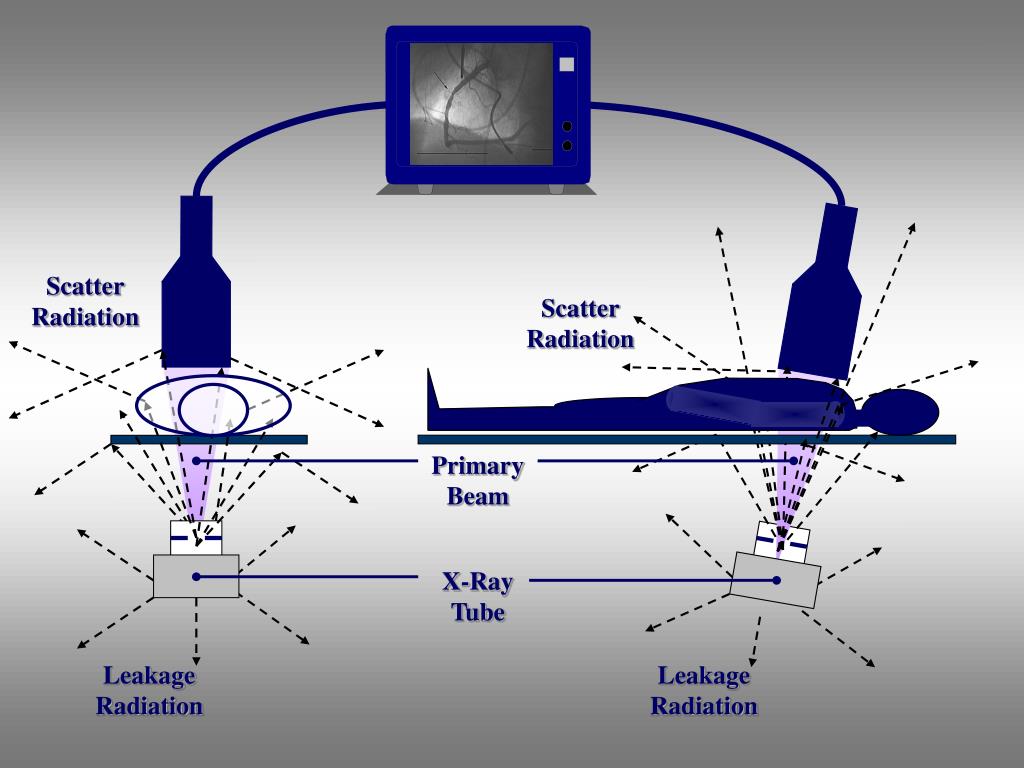
Values reported by integrated KAP/air kerma meters are air kerma values at a specified point in space, usually referred to as the interventional reference point (IRP). Patient entrance exposures are now reported as air kerma values and indicate the value that would be measured in a patient’s absence to eliminate effects due to backscatter in the patient. Air kerma is used to characterize the intensity of the x-ray beam 2 and has replaced the roentgen (R), the old unit of beam intensity. Most interventional equipment will report air kerma in milligray (mGy) as well as kerma-area product (KAP). For x-rays in air, the kerma is somewhat higher than the absorbed dose, as some of the released energy will be carried out of the small test volume as electron kinetic energy and will not contribute to the local dose.Īir kerma is the kerma in a small irradiated air volume. One Gy is equivalent to one joule (J) of energy per kilogram of matter. 1 The unit of measurement is the gray (Gy). For x-rays in tissue, this is numerically equivalent to the absorbed dose. This is the energy released from an x-ray beam per unit mass of a specified material in a small irradiated volume of matter (eg, air, soft tissue). Kinetic Energy Released in Matter (kerma) Common terms to clarify and add precision to our communication on matters regarding “dose” are reviewed herein. It is not comprehensive therefore, the references cited should be considered suggested reading for the motivated reader to learn more. The purpose of this article is to provide a “field guide” that can be used as a quick review prior to reading the rest of this issue of Endovascular Today and also as a quick reference in the future. Given these common uses, attention should be paid to the context, as this will often reveal the intent. In this situation, the ED is a more relevant parameter, although we are currently unable to estimate individual risks accurately. When patients or their families inquire about the dose from a procedure, they are more likely concerned about the risk of cancer induction, given the attention paid to this issue in the popular press.
#Scatter radiation skin
For complex procedures, we are concerned about the risk of adverse skin reactions, in which case the dose of interest is the PSD. We want to optimize dose and speak of it with regard to patients, staff, and operators, but what is it? “Dose” may mean absorbed dose or effective dose (ED), but it may also refer to peak skin dose (PSD) or even x-ray tube output. Interventionalists use the term “dose” on a daily basis and in a fairly casual fashion. The average interventionist may not use this terminology on a regular basis therefore, definitions and acronyms can become blurred, and terms can be used imprecisely. These results may be used for optimization of CBCTM systems, as well as for developing scatter correction methods.Radiation safety is an important topic for interventionists, and one of the most confusing aspects of this field is the variety of terms that are used. In general, the scatter to primary ratio (SPR) is strongly dependent on the breast size and air gap, and is only moderately dependent on the kVp setting and breast composition.

The influences of different air gaps, kVp settings, breast sizes and breast composition on the scatter primary ratio (SPR) and scatter profiles were examined. In this study, a GEANT4 based Monte Carlo simulation package (GATE) was used to investigate the scatter magnitude and its" distribution in CBCTM. In optimizing CBCTM systems, it is important to have knowledge of how different imaging parameters affect the recorded scatter within the image. Since the breast shape, geometry and image formation process are significantly different from conventional mammography, all system components and parameters such as target/filter combination, kVp range, source to image distance, detector design etc. One of the major challenges in CBCTM is to understand the characteristics of scatter radiation and to find ways to reduce or correct its degrading effects. Cone beam CT mammography (CBCTM) is an emerging breast imaging technology and is currently under intensive investigation.


 0 kommentar(er)
